
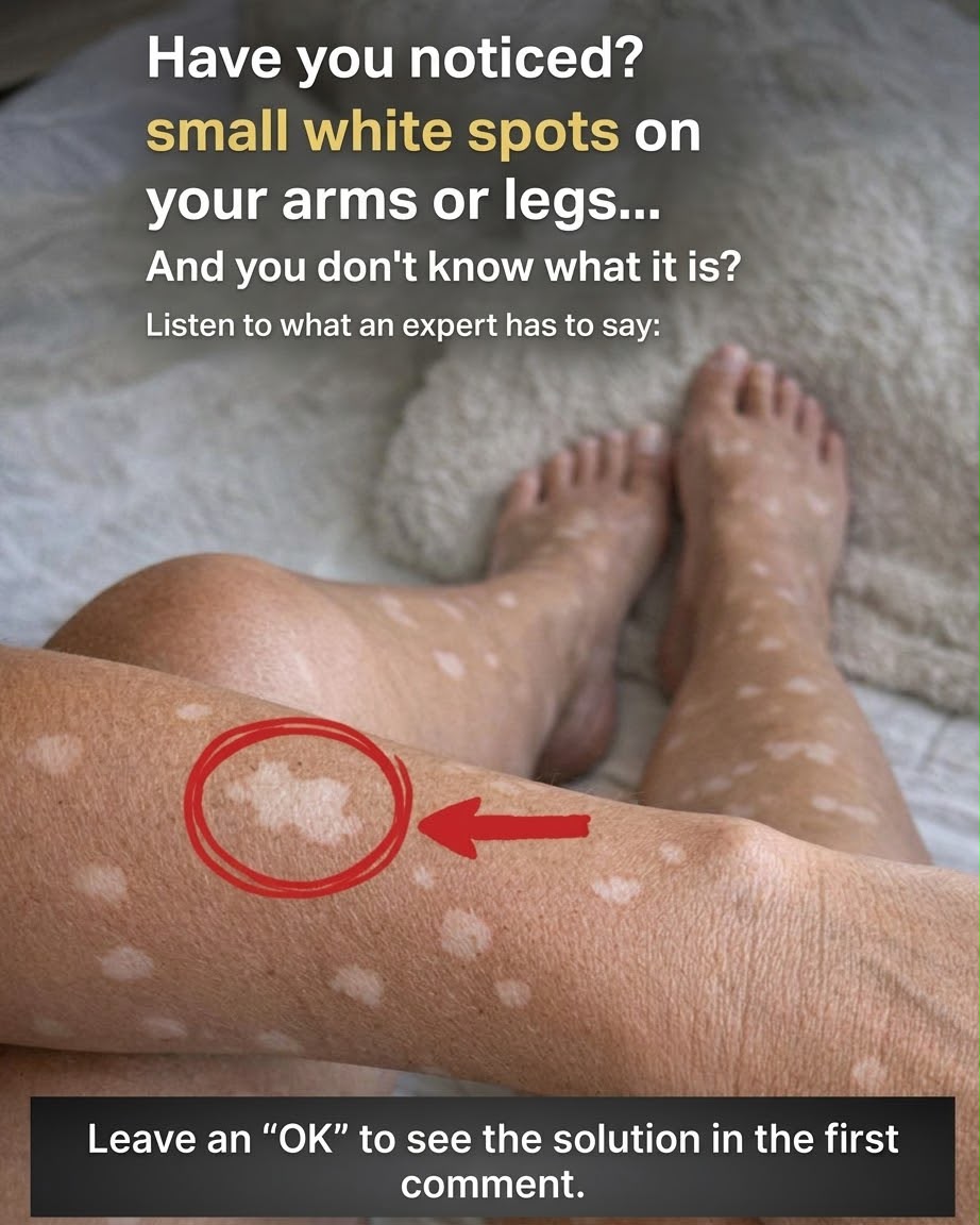
Have you ever noticed tiny, flat white spots or light patches appearing on your shins, forearms, or shoulders? Many adults discover these localized areas of pigment loss—known as hypopigmentation or depigmentation—as they get older or after spending time in the sun. While noticing sudden changes on your skin can be concerning, dermatologists emphasize that the vast majority of small white spots are completely harmless and tied to natural skin aging, sun exposure, or mild superficial skin conditions.
The human skin relies on specialized cells called melanocytes to produce melanin, the pigment responsible for giving skin its natural color and providing baseline protection against ultraviolet radiation. When these cells slow down pigment production, lose melanin density, or experience localized inflammation, smooth light or white patches emerge.
In this comprehensive guide, we will break down the primary medical causes for small white spots on your limbs, explore expert dermatological explanations, discuss over-the-counter and clinical management options, and examine how skin health intersects with preventive medical care and overall financial well-being.
The Most Common Causes of Small White Spots on Arms and Legs
Dermatologists categorize small white macules (flat spots) based on their distinct physical characteristics, texture, and underlying skin mechanisms.
1. Idiopathic Guttate Hypomelanosis (IGH)
By far the most common cause of tiny white spots on sun-exposed skin, Idiopathic Guttate Hypomelanosis (IGH) manifests as small, smooth, porcelain-white spots ranging from 1 to 10 millimeters in diameter.
Who It Affects: Most frequently seen in adults over the age of 40, particularly those with a history of long-term sun exposure.
Where It Appears: Primarily on the shins, lower legs, forearms, and upper shoulders.
The Cause: IGH is a completely benign process where localized melanocytes gradually decrease in number due to cumulative sun damage and natural aging.
2. Tinea Versicolor (Pityriasis Versicolor)
Tinea versicolor is a common, non-contagious fungal infection caused by an overgrowth of Malassezia, a yeast naturally present on human skin.
Appearance: Appears as small, slightly scaly patches that can look lighter (hypopigmented) or darker than surrounding skin, especially after tanning.
Key Trigger: Hot weather, humidity, oily skin, and excessive sweating encourage yeast proliferation, which secretes azelaic acid—a substance that temporarily blocks normal melanin synthesis.
3. Pityriasis Alba
Pityriasis alba is a mild, low-grade form of eczema or dermatitis frequently seen in children and young adults with sensitive or dry skin.
Appearance: Starts as slightly red, dry, or scaly patches that fade into soft, pale white spots with ill-defined borders.
Treatment: Responds well to gentle moisturizing creams, mild barrier repair lotions, and minimizing harsh soaps.
4. Vitiligo
Unlike IGH or pityriasis alba, vitiligo is a chronic autoimmune condition where the body’s immune system selectively destroys melanocytes.
Appearance: Results in larger, chalk-white, starkly defined depigmented patches that often present symmetrically on both sides of the body (such as both knees, wrists, or hands).
Progression: Patches can gradually enlarge over time and may involve hair follicles, turning overlying hair white.
Comparing Medical Conditions Causing White Spots
Understanding the subtle physical differences between skin conditions helps clarify when to monitor at home and when to consult a specialist.
Skin Condition Physical Appearance Texture Primary Underlying Cause
Idiopathic Guttate Hypomelanosis (IGH)
Small (1–5 mm), porcelain-white macules
Smooth, normal skin surface Cumulative sun exposure & natural aging
Tinea Versicolor Patchy, light-colored or reddish spots Slightly dry or fine powdery scale Skin yeast (Malassezia) overgrowth
Pityriasis Alba Pale, soft white patches with indistinct borders Dry, flaky, or mildly irritated Mild eczematous skin inflammation
Vitiligo
Stark chalk-white, symmetrical patches
Smooth, depigmented skin
Autoimmune destruction of melanocytes
What Dermatologists Say: Management and Treatments
While harmless conditions like IGH do not require medical intervention, various clinical dermatological treatments can help improve skin tone uniformity for individuals seeking cosmetic refinement.
Retinoids and Topical Therapies
Prescription topical retinoids (such as tretinoin) can stimulate epidermal cell turnover and encourage localized melanocyte activity over several months of consistent use. For conditions like pityriasis alba or eczema-related lightness, mild topical anti-inflammatory creams restore the skin barrier.
Antifungal Wash Strategies for Tinea Versicolor
If yeast overgrowth is diagnosed via skin scraping or Wood’s lamp examination, dermatologists typically prescribe topical antifungal washes containing ketoconazole, selenium sulfide, or zinc pyrithione. Applying these cleansers for a few minutes before rinsing clears the fungal overgrowth, allowing normal pigment to return gradually as the skin tans or renews itself.
Advanced Light Therapy (Phototherapy)
For significant depigmentation conditions like vitiligo, narrow-band UVB phototherapy or targeted excimer laser treatments are used in clinical settings to stimulate dormant melanocytes deep within hair follicles, promoting repigmentation.
Preventive Skin Care: Protecting Your Skin Barrier
Whether you are managing existing white spots or preventing new sun-induced spots from forming, establishing an effective daily skin protection routine is essential.
Broad-Spectrum Daily Sunscreen
Because UV radiation accelerates melanocyte depletion and damages skin DNA, applying a broad-spectrum, water-resistant sunscreen with SPF 30 or higher to exposed arms and legs is crucial year-round. Sun protection prevents surrounding skin from darkening dramatically, which minimizes the contrast between white spots and normal skin tone.
Barrier-Restoring Moisturization
Applying rich emollients containing ceramides, hyaluronic acid, and niacinamide immediately after showering seals in moisture, reduces dry flaking, and calms mild skin inflammation that can trigger post-inflammatory hypopigmentation.
Connecting Skin Health to Medical Insurance and Financial Peace of Mind
Addressing skin changes proactively is a key aspect of maintaining overall health and preventing unnecessary anxiety.
Maximizing Preventive Healthcare and Insurance Benefits
When you notice new skin lesions, spots, or pigment shifts, scheduling an annual full-body skin exam with a board-certified dermatologist ensures that serious conditions—such as early-stage skin cancers—are ruled out. Under most standard health insurance policies, routine annual skin screenings and diagnostic consultations are covered medical benefits after applicable copays or deductibles.
Financial Planning and Life Security
Staying proactive with routine medical checkups prevents minor skin conditions from developing into complex health issues. Maintaining optimal wellness helps protect your personal finance goals, safeguarding your ongoing ability to work, build savings, and fund your family’s long-term investment and retirement strategy. Ensuring adequate health, disability, and life insurance coverage provides complete financial security for life’s uncertainties.
Fascinating Facts About Skin Pigmentation
The World’s Largest Organ: The skin accounts for roughly 16% of total human body weight and continuously sheds roughly 30,000 to 40,000 dead skin cells every single minute.
Melanin as a Natural Shield: Melanin acts as an internal biological umbrella, forming protective caps over the DNA inside skin cell nuclei to absorb and scatter incoming UV rays.
Sun Memory: The skin “remembers” UV exposure; sun spots, freckles, and IGH white spots often reflect sunburns and sun exposure that occurred 10 to 20 years earlier in life.
Melanocyte Density: Surprisingly, human beings of all skin tones possess roughly the same number of melanocytes per square inch of skin. Differences in skin tone stem from the size, distribution, and activity levels of the melanin packages (melanosomes) these cells produce.
Conclusion: Understanding and Caring for Your Skin
Discovering small white spots on your arms or legs is a very common experience that rarely indicates a dangerous medical condition. Whether caused by natural sun-related changes like IGH, mild yeast imbalances, or dry skin, understanding the root cause empowers you to care for your skin effectively.
By practicing consistent daily sun protection, keeping your skin thoroughly hydrated, and utilizing your health insurance benefits for annual dermatological exams, you can maintain healthy, radiant skin and peace of mind at every age.
Frequently Asked Questions
Are small white spots on arms and legs a sign of skin cancer?
No, small white spots like Idiopathic Guttate Hypomelanosis (IGH) or pityriasis alba are completely benign and are not precancerous or cancerous. However, any new, rapidly changing, or irregular skin growth should always be evaluated by a dermatologist to ensure proper diagnosis.
Can diet cause white spots on the skin?
While mild nutritional deficiencies in Vitamin D, Vitamin B12, or zinc can sometimes impact overall skin health and healing, small white spots on the limbs are predominantly caused by sun exposure, skin aging, local inflammation, or yeast overgrowth rather than specific dietary factors.
Will IGH white spots ever go away naturally?
White spots caused by IGH typically do not disappear on their own because the local pigment-producing cells have decreased. However, keeping the skin well-moisturized, applying daily sunscreen to prevent new spots, and using cosmetic topical treatments can help make them far less noticeable.
How can I tell if my white spots are vitiligo or sun spots?
Sun spots (IGH) are usually tiny (1–5 mm), isolated, smooth, and appear mostly on lower legs and arms. Vitiligo typically forms larger, stark white patches that often appear symmetrically on both sides of the body (such as both elbows, wrists, or knees) and can spread over time.
Can I tan to blend white spots with the rest of my skin?
Tanning is not recommended. Tanning darkens the surrounding healthy skin, which actually increases the visual contrast and makes white spots stand out more prominently. Furthermore, UV exposure accelerates skin aging and increases the risk of developing additional IGH spots.
